2026 · Hemodynamics & imaging
Systematic machine learning approach for cerebral aneurysm feature selection and rupture status classification
Kofman LA, Ludwig CG, Lesha E, Lauric A, Malek AM.
J Clin Neurosci 144:111803, 2026.
PMID 41370993 · DOI 10.1016/j.jocn.2025.111803
In plain language
Doctors decide whether to treat an unruptured brain aneurysm partly by measuring its size and shape by hand. This study tested seven machine learning methods on the 3D angiograms of 678 aneurysms, 229 of which had ruptured, to see whether a computer could tell ruptured from unruptured aneurysms based on their shape and location.
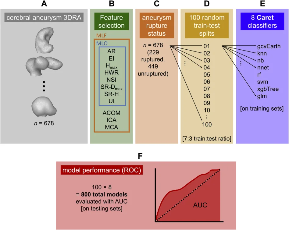
The best models performed well, with an area under the curve of about 0.80 to 0.83 (1.0 would be perfect), and did better than standard statistical methods. Measures of how irregular or elongated an aneurysm is mattered more than its overall size.
This automated approach could help make rupture risk assessment more consistent.
A plain-language summary for patients and families. It is not medical advice, and individual results vary.
Abstract
Background
Current methods of intracranial aneurysm rupture risk assessment in the clinical setting depend on user measurements of morphological factors. Recently, machine learning (ML) has been proposed to assist in stratification of aneurysmal rupture risk. While numerous approaches have been taken, most rely on small sample sizes, restrict to specific anatomical locations, or manually balance ruptured and unruptured samples to enhance classification. By using 7 ML classifiers on a retrospective cohort of consecutive aneurysms distributed throughout the cranial vasculature, we present a computational tool that effectively classifies aneurysmal rupture status on a large, generalizable dataset.
Methods
Three-dimensional angiograms of patient cerebral aneurysms (n = 678; 229 ruptured and 449 unruptured aneurysms) enabled computation of 21 aneurysm features. Two datasets were used: morphological features only (MFO), and morphological plus 15 locational features (MLF). After normalization of numerical features and 7:3 training-testing splitting, for feature selection, a multivariate elastic network regression model was trained via 5-fold cross validation. Features were selected on the testing set. For classification, random 7:3 training-testing splitting was performed 100 times independently on each stratus. 8 classifiers (7 ML models and 1 generalized linear model; GLM) were used to assess rupture status. Area under the curve (AUC), sensitivity (SENS), and specificity (SPEC) were evaluated on testing sets. To elucidate the contribution of each feature to aneurysm rupture status assessment, feature importance was calculated for each optimal classifier.
Results
On the MFO dataset, multiple adaptive regression splines (MARSMFO) exhibited the greatest performance (AUCmean = 0.797, SENSmean = 0.777, SPECmean = 0.728), with AUC also significant compared to GLM and three other ML classifiers. Aspect ratio (AR) and undulation index (UI) were the strongest factors. On the MLF dataset, neural networks (NNETMLF) achieved the greatest performance (AUCmean = 0.825, SENSmean = 0.803, SPECmean = 0.738), followed by random forests (RFMLF; AUCmean = 0.825, SENSmean = 0.847, SPECmean = 0.710). For NNETMLF, AUC was found significant compared to GLM and three other ML classifiers. In neural networks, non-sphericity index (NSI), middle cerebral artery location, and AR were identified as the strongest contributors to rupture status.
Conclusion
We propose a systematic, user-independent approach to ML aneurysm rupture status classification. MARSMFO and NNETMLF exhibited the highest AUCs and outperform the traditional GLM, suggesting the utility of ML to assess intracranial rupture risk status. Morphological features such as NSI, UI, and AR were selected over maximal diameter, volume, and area. These factors may empower clinicians to accurately assess rupture risk, and they should be further studied.
Abstract as indexed in PubMed (PMID 41370993). Copyright the publisher.
Cite this paper
Kofman LA, Ludwig CG, Lesha E, Lauric A, Malek AM. Systematic machine learning approach for cerebral aneurysm feature selection and rupture status classification. J Clin Neurosci 144:111803. 2026. doi:10.1016/j.jocn.2025.111803 PMID: 41370993.