2014 · Hemodynamics & imaging
Superior performance of cone-beam CT angiography in characterization of intracranial atherosclerosis
Safain MG, Rahal JP, Patel S, Lauric A, Feldmann E, Malek AM.
J Neurosurg 121(2):441-9, 2014.
PMID 24949672 · DOI 10.3171/2014.5.JNS132201
In plain language
Intracranial atherosclerotic disease is a buildup of fatty plaque inside the arteries of the brain, and it carries a high risk of stroke. Doctors have mainly judged it by how much the artery is narrowed, but the shape and surface features of the plaque itself are increasingly thought to matter.
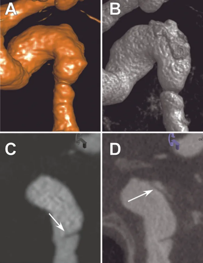
This first report on the topic looked at 18 patients who had catheter angiograms between August 2009 and July 2012. Each was imaged with three methods: standard 2D angiography, 3D rotational angiography, and ultra-high-resolution cone-beam CT angiography. The two 3D methods measured the narrowing similarly, while both differed from standard 2D angiography. Cone-beam CT showed plaque features best, such as ulcers, calcium deposits, tears and spiky edges, revealing a new plaque feature in 61% of patients compared with 2D angiography and in 50% compared with 3D rotational angiography.
Further studies are needed to see whether this extra detail improves treatment decisions.
A plain-language summary for patients and families. It is not medical advice, and individual results vary.
Abstract
Object
Intracranial atherosclerotic disease (ICAD) carries a high risk of stroke. Evaluation of ICAD has focused on assessing the absolute degree of stenosis, although plaque morphology has recently demonstrated increasing relevance. The authors provide the first report of the use of ultra-high-resolution C-arm cone-beam CT angiography (CBCT-A) in the evaluation of vessel stenosis as well as plaque morphology.
Methods
Between August 2009 and July 2012, CBCT-A was used in all patients with ICAD who underwent catheter-based angiography at the authors' institution (n = 18). Lesions were evaluated for maximum degree of stenosis as well as plaque morphological characteristics (ulcerated, calcified, dissected, or spiculated) via digital subtraction angiography (DSA), 3D-rotational angiography (3DRA), and CBCT-A. The different imaging modalities were compared in their assessment of absolute stenosis as well as their ability to resolve different plaque morphologies.
Results
Lesions were found to have similar degrees of stenosis when utilizing CBCT-A compared with 3DRA, but both 3DRA and CBCT-A differed from DSA in their assessment of the absolute degree of stenosis. CBCT-A provided the most detailed resolution of plaque morphology, identifying a new plaque characteristic in 61% of patients (n = 11) when compared with DSA and 50% (n = 9) when compared with 3DRA. CBCT-A identified all lesion characteristics visualized on DSA and 3DRA.
Conclusions
CBCT-A provides detailed spatial resolution of plaque morphology and may add to DSA and 3DRA in the evaluation of ICAD. Further prospective study is warranted to determine any benefit CBCTA-A may provide in clinical decision making and risk stratification over existing conventional imaging modalities.
Abstract as indexed in PubMed (PMID 24949672). Copyright the publisher.
Cite this paper
Safain MG, Rahal JP, Patel S, Lauric A, Feldmann E, Malek AM. Superior performance of cone-beam CT angiography in characterization of intracranial atherosclerosis. J Neurosurg 121(2):441-9. 2014. doi:10.3171/2014.5.JNS132201 PMID: 24949672.