2013 · Devices & clinical
Successful detection of embologenic ulceration in a symptomatic non-hemodynamic intracranial stenosis using C-arm cone beam CT
Heller RS, Malek AM.
J Neurointerv Surg 5(2):e3, 2013.
PMID 22266793 · DOI 10.1136/neurintsurg-2011-010206
In plain language
Narrowing of arteries inside the skull from fatty plaque buildup is usually examined with catheter angiography, an X-ray movie of the vessels. This case report describes a 73-year-old man with worsening mini-strokes (brief episodes of right arm tingling and hand weakness) despite taking two blood-thinning medications. Standard angiography showed only a smooth, mild narrowing of the left carotid artery near the skull base.
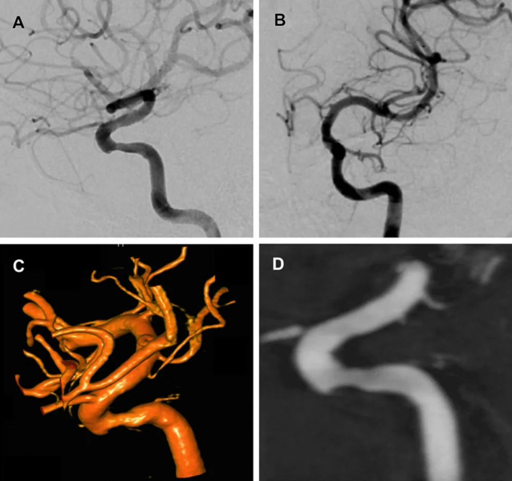
Because the image did not match his symptoms, the team used C-arm cone beam CT, a higher-resolution 3D scan done in the angiography room. It revealed a broken, ulcerated, calcified plaque with a small tear and an overlying clot. He was treated with anticoagulation and then a balloon-mounted stent, and his symptoms resolved with the artery still open at 1 year.
Such imaging may help sort out uncertain plaques that ordinary angiography misses.
A plain-language summary for patients and families. It is not medical advice, and individual results vary.
Abstract
Objective
The gold standard for the diagnosis of intracranial atherosclerosis remains catheter based digital subtraction angiography (DSA). A symptomatic hemodynamically non-significant intracranial atherosclerotic lesion is described, whose underlying embologenic characteristics were not detectable by either DSA or three-dimensional rotational angiography (3DRA) but fully realized by C-arm cone beam CT (CBCT) angiography.
Clinical presentation
A 73-year-old man presented with crescendo transient ischemic attacks consisting of right arm tingling and hand weakness despite long term dual antiplatelet therapy with aspirin and clopidogrel for coronary artery stent. DSA and 3DRA demonstrated a smooth benign appearing left cavernous internal carotid stenosis of <50%.
Intervention
Given the incongruence of the lesion with the patient's symptoms and lack of response to aggressive medical treatment, a decision was made to obtain higher resolution imaging. CBCT angiography was obtained with injection at two contrast dilutions, which uncovered an underlying ruptured ulcerated calcific plaque with a small dissective component and overlying thrombus. The lesion was treated with anticoagulation followed by balloon mounted stent angioplasty, with symptom resolution and maintained patency at the 1 year follow-up.
Conclusion
The superior spatial resolution and dynamic range characteristics of CBCT angiography provide added clinical utility in disambiguation of questionable intracranial atherosclerotic lesions which may be missed by conventional planar and rotational angiography. The additional information provided by CBCT angiography could be useful in lesion risk stratification and help refine indications for intracranial stent angioplasty given its recent documented shortcomings vis a vis medical management.
Abstract as indexed in PubMed (PMID 22266793). Copyright the publisher.
Cite this paper
Heller RS, Malek AM. Successful detection of embologenic ulceration in a symptomatic non-hemodynamic intracranial stenosis using C-arm cone beam CT. J Neurointerv Surg 5(2):e3. 2013. doi:10.1136/neurintsurg-2011-010206 PMID: 22266793.