2018 · Hemodynamics & imaging
Critical role of angiographic acquisition modality and reconstruction on morphometric and haemodynamic analysis of intracranial aneurysms
Lauric A, Hippelheuser JE, Malek AM.
J Neurointerv Surg 10(9):911-915, 2018.
PMID 29352062 · DOI 10.1136/neurintsurg-2017-013677
In plain language
Researchers often use 3D images of aneurysms to measure their shape and to run computer simulations of blood flow. Results vary between studies, however, and part of the reason may be the way the images are taken and processed.
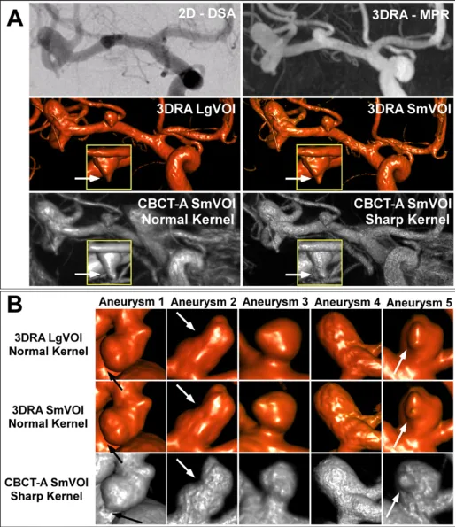
Four patients with five aneurysms had both 3D rotational angiography and high-resolution cone-beam CT angiography during the same session. The authors rebuilt six models of each aneurysm, varying the imaging method, the image detail (voxel size) and the processing filter, called a kernel. These choices significantly changed measurements such as aneurysm size and neck width, as well as wall shear stress, the rubbing force of blood on the wall. Cone-beam CT gave shapes differing by up to 24% and flow results differing by up to 97% compared with 3D rotational angiography.
Such differences in technique can hide real differences between patients and make it hard to compare studies from different centers.
A plain-language summary for patients and families. It is not medical advice, and individual results vary.
Abstract
Background
Subtracted 3-D rotational angiography (3DRA) and cone-beam computed tomography angiography (CBCT-A) are often used in assessing cerebral aneurysm shape and haemodynamic profile. We sought to evaluate the effect of imaging modality, reconstruction parameters, and kernel selection on patient-derived aneurysm morphology and computational fluid dynamic (CFD) analysis to assess its potential contribution to inter-study variability.
Methods
Four patients (five aneurysms) underwent concurrent 3DRA followed by high-resolution CBCT-A. Six models were reconstructed per aneurysm: 3DRA reconstructed with 0.28 and 0.14 mm voxel sizes (large and small volume of interest (VOI) respectively), and two kernel types (normal/smooth). CBCT-A was reconstructed over small VOI using normal/sharp kernel. Maximal dome dimension, neck diameter and dome/neck ratio were evaluated in 3D. Wall shear stress (WSS) magnitude was evaluated on the entire aneurysm dome and in the 5% dome areas covered by lowest (LWSS) and highest (HWSS) WSS. Parameters were evaluated with pairwise t-test analysis.
Results
Smaller VOI reconstructions resulted in smaller Dmax (P value=0.03) and Dmax/neck (P value=0.006) and in larger LWSS (P value=0.03). Use of sharp kernel led to narrower neck (P value=0.04) and higher Dmax/neck values (P value=0.02). CBCT-A resulted in statistically different aneurysm shape (up to 24% difference) and haemodynamics (up to 97% difference) compared with 3DRA.
Conclusion
The choice of catheter 3D angiographic modality and reconstruction kernel has a critical impact on derived aneurysm morphological and haemodynamic analysis. The resultant variability can confound and obscure underlying differences within patient populations and between studies performed at different centres using divergent techniques, compromising the accuracy of quantitative aneurysm analysis.
Abstract as indexed in PubMed (PMID 29352062). Copyright the publisher.
Cite this paper
Lauric A, Hippelheuser JE, Malek AM. Critical role of angiographic acquisition modality and reconstruction on morphometric and haemodynamic analysis of intracranial aneurysms. J Neurointerv Surg 10(9):911-915. 2018. doi:10.1136/neurintsurg-2017-013677 PMID: 29352062.