2013 · Devices & clinical
Neuroform intracranial stenting for aneurysms using simple and multi-stent technique is associated with low risk of magnetic resonance diffusion-weighted imaging lesions
Heller RS, Dandamudi V, Calnan D, Malek AM.
Neurosurgery 73(4):582-90; discussion 590-1, 2013.
PMID 23787881 · DOI 10.1227/NEU.0000000000000053
In plain language
Small clots can break off during stent-assisted aneurysm treatment and cause tiny strokes that show up on a sensitive MRI scan called diffusion-weighted imaging. Little was known about how often this happens with the Neuroform stent, which has an open-cell design.
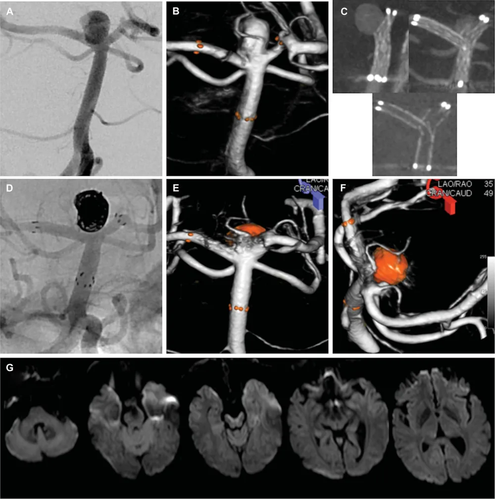
This study reviewed 76 patients who received 97 Neuroform stents and had an MRI within 48 hours. New spots were found in 7 of 78 procedures (9.0%), and none caused a permanent deficit at discharge. Using multiple stents, including the Y-shaped two-stent technique, did not lead to any new spots, nor did treatment after aneurysm bleeding. The only related factor was an aneurysm on the side wall of a vessel rather than at a branch point.
The Neuroform stent carried a low risk of these small clots.
A plain-language summary for patients and families. It is not medical advice, and individual results vary.
Abstract
Background
Detection of procedural thromboembolism by diffusion-weighted magnetic resonance imaging (MR-DWI+) can help identify and mitigate endovascular risk factors. Data remain scant on procedural MR-DWI+ following the use of the Neuroform open-cell design stent in aneurysm embolization.
Objective
We sought to evaluate the incidence of MR-DWI+ in Neuroform simple and multi-stent construct stenting for intracranial aneurysms in an attempt to delineate baseline risk and identify possible associated procedural factors.
Methods
Seventy-six patients receiving 97 Neuroform stents in the treatment of intracranial aneurysm were identified from a prospective database and eligible for inclusion in the study. Diffusion-weighted magnetic resonance imaging (MR-DWI) was obtained in all patients within 48 hours of the procedure and reviewed for the presence of MR-DWI+ with patient records reviewed for analysis of factors predisposing to these lesions.
Results
Patients were treated with single-stent Neuroform constructs in 57 cases (73%) and multi-stent Neuroform constructs in 21 cases (27%). Y-stent technique was used in 16 cases. MR-DWI+ was identified in 7 of 78 cases (9.0%), with MR-DWI+ in 0 of 10 subarachnoid hemorrhage cases. No MR-DWI lesions led to a permanent neurological deficit at discharge. There was no MR-DWI+ in patients treated with Y-stenting or multi-stent Neuroform constructs. The only factor associated with ipsilateral MR-DWI+ was target aneurysm location on an arterial sidewall over bifurcation (P = .01).
Conclusion
The Neuroform stent carries a very low risk of MR-DWI+ compared with its closed-cell design counterpart. Subarachnoid hemorrhage and deployment of multiple stents in the same anatomical region in configurations such as the Y-stent construct did not increase the risk of acute procedural thromboembolism.
Abstract as indexed in PubMed (PMID 23787881). Copyright the publisher.
Cite this paper
Heller RS, Dandamudi V, Calnan D, Malek AM. Neuroform intracranial stenting for aneurysms using simple and multi-stent technique is associated with low risk of magnetic resonance diffusion-weighted imaging lesions. Neurosurgery 73(4):582-90; discussion 590-1. 2013. doi:10.1227/NEU.0000000000000053 PMID: 23787881.