2011 · Devices & clinical
Crescent sign on magnetic resonance angiography revealing incomplete stent apposition: correlation with diffusion-weighted changes in stent-mediated coil embolization of aneurysms
Heller RS, Miele WR, Do-Dai DD, Malek AM.
J Neurosurg 115(3):624-32, 2011.
PMID 21619405 · DOI 10.3171/2011.4.JNS102050
In plain language
Stents, small mesh tubes, are sometimes placed in an artery to hold coils inside a wide brain aneurysm. This study asked how closely these stents press against the vessel wall and whether gaps matter, using MRI scans in 58 patients treated with either the Enterprise (closed-cell) or Neuroform (open-cell) stent.
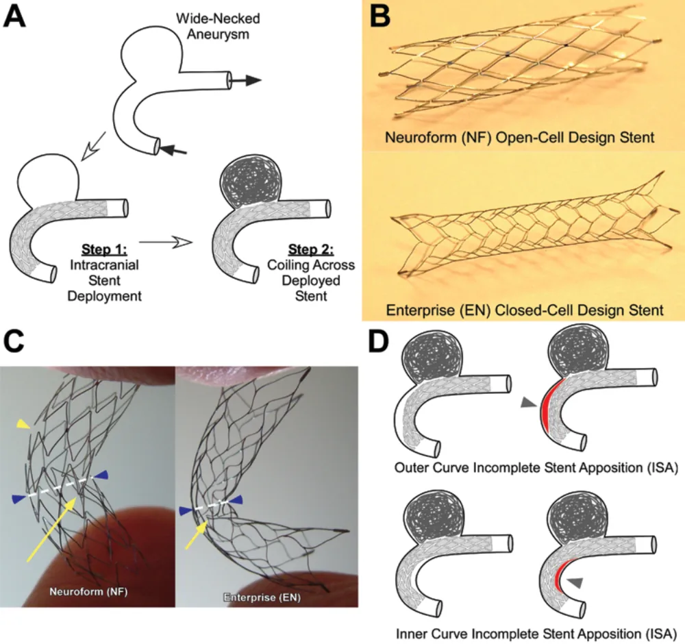
The scans showed a half-moon pattern, named the crescent sign, reflecting blood flowing outside the stent where it did not touch the wall. It appeared in 18 of 33 Enterprise cases and 0 of 25 Neuroform cases, more often in the curving internal carotid artery. The crescent sign strongly predicted small new spots on diffusion MRI, a scan that detects tiny areas of stroke, on the treated side.
Poor stent contact may therefore be linked to clot-related complications.
A plain-language summary for patients and families. It is not medical advice, and individual results vary.
Abstract
Object
Few data are available on how closely stents appose the luminal vessel wall in stent-mediated coil embolization of intracranial aneurysms and on the effect of incomplete stent apposition on procedural thromboembolic complications.
Methods
Postprocedural 3-T MR diffusion-weighted imaging and time-of-flight angiography were obtained in 58 patients undergoing stent-mediated coil embolization of aneurysms using the Enterprise closed-cell and Neuroform open-cell self-expanding intracranial microstents.
Results
A distinctive semilunar signal pattern, identified using 3-T MR angiography, represented flow outside the confines of the stent struts in patients in whom Enterprise but not Neuroform devices were used. This pattern, designated as the crescent sign, was confirmed to correspond to incomplete stent apposition by use of high-resolution angiographic flat-panel CT scanning revealing flow ingress into and egress out of the isolated luminal wedge. The presence of the crescent sign was seen in 18 of 33 Enterprise-treated but in 0 of 25 Neuroform-treated cases, and was more likely in stents delivered in the tortuous internal carotid artery (p = 0.034). The crescent sign was strongly predictive of ipsilateral postprocedural lesions seen on diffusion-weighted imaging in the entire population (OR 18, 95% CI 4.33-74.8; p < 0.0001). In the Enterprise stent subset, ipsilateral lesions were detected on diffusion-weighted imaging in 15 (45%) of 33 cases; the crescent sign was seen in 12 (80%) of 15 patients with ipsilateral lesions on diffusion-weighted imaging, but in only 6 of 18 patients without lesions (OR 8, 95% CI 1.61-39.6; p = 0.006).
Conclusions
Incomplete stent apposition is detectable on 3-T MR angiography as a crescent sign, and was found to be highly prevalent in Enterprise closed-cell design stents used to assist coil embolization of aneurysms. Incomplete stent apposition was also associated with periprocedural ipsilateral hyperintense lesions on diffusion-weighted imaging. These results identify an association between incomplete stent apposition and thromboembolic complications in stent-mediated coil embolization of intracranial aneurysms.
Abstract as indexed in PubMed (PMID 21619405). Copyright the publisher.
Cite this paper
Heller RS, Miele WR, Do-Dai DD, Malek AM. Crescent sign on magnetic resonance angiography revealing incomplete stent apposition: correlation with diffusion-weighted changes in stent-mediated coil embolization of aneurysms. J Neurosurg 115(3):624-32. 2011. doi:10.3171/2011.4.JNS102050 PMID: 21619405.