2010 · Devices & clinical
Intraoperative use of indocyanine green fluorescence videography for resection of a spinal cord hemangioblastoma
Hwang SW, Malek AM, Schapiro R, Wu JK.
Neurosurgery 67(3 Suppl Operative):ons300-3; discussion ons303, 2010.
PMID 20679920 · DOI 10.1227/01.NEU.0000383876.72704.7B
In plain language
Indocyanine green is a fluorescent dye that, once injected into the bloodstream, lights up blood vessels and tissue under a special camera during surgery. It has mostly been used in brain aneurysm operations, with little reported use in spine tumor surgery.
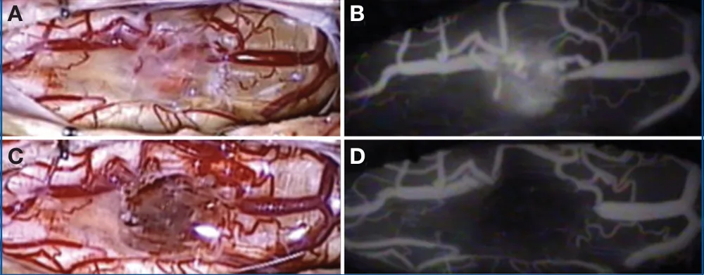
This case report describes a 49-year-old woman with a hemangioblastoma, a tumor rich in blood vessels, in the upper spinal cord at the neck. It remained after earlier treatment elsewhere and was causing worsening symptoms. During surgery, the dye revealed part of the tumor hidden under thick, cloudy tissue that could not be seen through the microscope. Imaging right after removal suggested the tumor was fully removed, which a later MRI confirmed.
The authors find the dye especially useful for leftover or recurrent tumors hidden by scar-like adhesions.
A plain-language summary for patients and families. It is not medical advice, and individual results vary.
Abstract
Background and importance
Indocyanine green (ICG) fluorescence videography has been recently applied to the neurosurgical field, mostly in the management of cerebral aneurysms, but has had limited description in the subspecialty of spine or oncological neurosurgery. We describe a novel application of this previously defined surgical tool to assist in the resection of a residual spinal cord hemangioblastoma.
Clinical presentation
Our patient is a 49-year-old woman with a residual symptomatic cervical hemangioblastoma that was previously embolized and resected at another institution. After initial symptomatic improvement, she returned with progressive symptoms, increasing radiographic spinal cord edema, and a residual lesion at the level of C1. We resected the remaining tumor with the adjuvant use of ICG fluorescence videography. Intraoperative injection of ICG clearly identified a component of the tumor underlying adhesive, opaque arachnoid that was not visualized by direct microscopy. Immediate postresection ICG videography suggested a complete resection was achieved which was later corroborated by postoperative magnetic resonance imaging.
Conclusion
The adjuvant use of ICG videography is a useful surgical tool that permits greater visualization of the complete extent of the lesion, particularly in managing recurrent or residual lesions obscured by adhesions.
Abstract as indexed in PubMed (PMID 20679920). Copyright the publisher.
Cite this paper
Hwang SW, Malek AM, Schapiro R, Wu JK. Intraoperative use of indocyanine green fluorescence videography for resection of a spinal cord hemangioblastoma. Neurosurgery 67(3 Suppl Operative):ons300-3; discussion ons303. 2010. doi:10.1227/01.NEU.0000383876.72704.7B PMID: 20679920.