2013 · Hemodynamics & imaging
Cerebral aneurysm wall thickness analysis using intraoperative microscopy: effect of size and gender on thin translucent regions
Kadasi LM, Dent WC, Malek AM.
J Neurointerv Surg 5(3):201-6, 2013.
PMID 22387724 · DOI 10.1136/neurintsurg-2012-010285
In plain language
The thickness of an aneurysm's wall is poorly understood but may relate to how aneurysms grow, rupture and respond to treatment. During open surgery, the surgical microscope lets surgeons see that some areas of the wall are thin enough to be see-through.
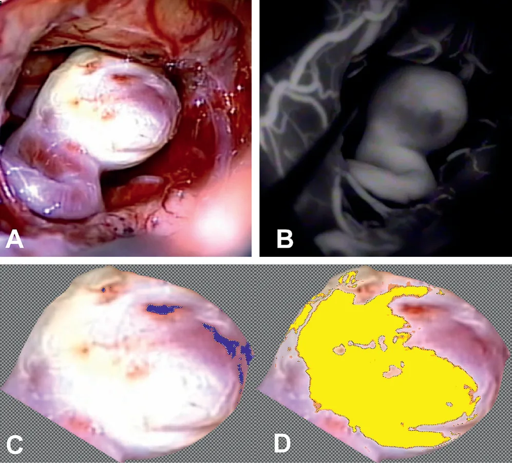
This study reviewed surgical video of 54 unruptured aneurysms and measured how much of each dome was very thin and translucent, intermediate, or thick, based on color. On average, domes were 27% very thin, 65% intermediate and 8% thick. Smaller aneurysms (under 7 millimeters) had more very thin tissue and less thick tissue, and aneurysms in women had more very thin tissue. Age, smoking and location made no difference.
The authors conclude that wall thickness varies greatly across the dome and should be included in future studies of rupture risk.
A plain-language summary for patients and families. It is not medical advice, and individual results vary.
Abstract
Objective
Wall thickness is a poorly documented characteristic of cerebral aneurysms which may provide insight into adaptive aneurysmal growth, aneurysm rupture risk and response to endovascular treatment. The distribution of aneurysm wall thickness, as observed by intraoperative video microscopy, is described.
Methods
54 unruptured saccular cerebral aneurysms were selected based on the availability of intraoperative video obtained from patients undergoing microsurgical clipping. Aneurysms were assessed for the distribution of wall thickness based on color translucence and quantitation of pixel values at superthin translucent, intermediate and thick regions of the dome. The data were analyzed with respect to aneurysm morphology, location and associated demographic factors.
Results
The mean proportions of tissue characteristic among all domes analyzed were found to be 27% superthin, 65% intermediate, and 8% thick. Smaller aneurysms having a maximal dimension Dmax <7 mm had a higher proportion of superthin tissue (p=0.003) and lower thick tissue (p=0.001) content. Female gender was associated with a significantly higher proportion of superthin tissue at the aneurysm dome (p=0.038), with no statistical dependence seen with patient age, smoking status or anatomical location.
Conclusion
The dome of unruptured aneurysms is a highly heterogeneous region with areas of variable thickness that appear to be intimately related to the process of aneurysm development. This inconstant property affects wall tensile stress, may play a role in aneurysm pathogenesis and focal rupture, and should be incorporated into future analyses of aneurysm rupture risk and mechanics.
Abstract as indexed in PubMed (PMID 22387724). Copyright the publisher.
Cite this paper
Kadasi LM, Dent WC, Malek AM. Cerebral aneurysm wall thickness analysis using intraoperative microscopy: effect of size and gender on thin translucent regions. J Neurointerv Surg 5(3):201-6. 2013. doi:10.1136/neurintsurg-2012-010285 PMID: 22387724.