2013 · Hemodynamics & imaging
Colocalization of thin-walled dome regions with low hemodynamic wall shear stress in unruptured cerebral aneurysms
Kadasi LM, Dent WC, Malek AM.
J Neurosurg 119(1):172-9, 2013.
PMID 23540271 · DOI 10.3171/2013.2.JNS12968
In plain language
Wall shear stress, the rubbing force of blood flowing along a vessel wall, affects how the cells lining the vessel behave and has been suspected in aneurysm rupture. This study asked whether thin spots on an aneurysm's wall line up with areas of low force.
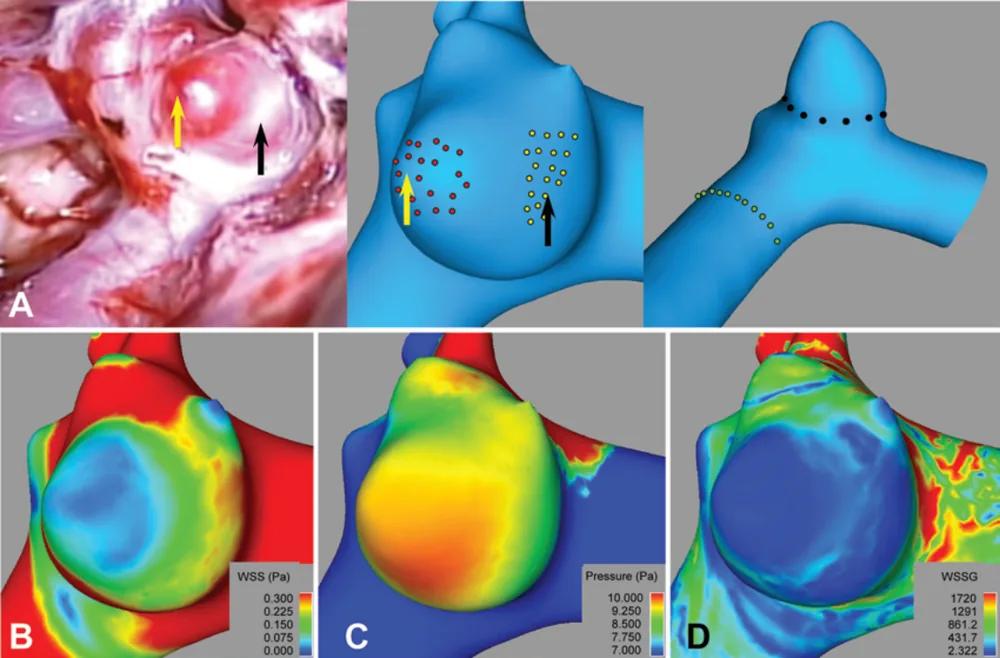
Computer flow simulations were done for 16 unruptured aneurysms in 14 patients, and the results were matched to views from the surgical microscope, where thin areas look translucent. Thin areas had much lower wall shear stress than nearby normal-thickness areas (0.381 Pa vs 0.816 Pa). Lower force and higher local pressure together identified thin areas well, with an area under the curve of 0.80 (1.0 would be perfect).
This suggests that sluggish flow may signal cells in the wall in ways that lead to thinning.
A plain-language summary for patients and families. It is not medical advice, and individual results vary.
Abstract
Object
Wall shear stress (WSS) plays a role in regulating endothelial function and has been suspected in cerebral aneurysm rupture. The aim of this study was to evaluate the spatial relationship between localized thinning of the aneurysm dome and estimated hemodynamic factors, hypothesizing that a low WSS would correlate with aneurysm wall degeneration.
Methods
Steady-state computational fluid dynamics analysis was performed on 16 aneurysms in 14 patients based on rotational angiographic volumes to derive maps of WSS, its spatial gradient (WSSG), and pressure. Local dome thickness was estimated categorically based on tissue translucency from high-resolution intraoperative microscopy findings. Each computational model was oriented to match the corresponding intraoperative view and numerically sampled in thin and normal adjacent dome regions, with controls at the neck and parent vessel. The pressure differential was computed as the difference between aneurysm dome points and the mean neck pressure. Pulsatile time-dependent confirmatory analysis was carried out in 7 patients.
Results
Matched-pair analysis revealed significantly lower levels of WSS (0.381 Pa vs 0.816 Pa; p<0.0001) in thin-walled dome areas than in adjacent baseline thickness regions. Similarly, log WSSG and log WSS×WSSG were both lower in thin regions (both p<0.0001); multivariate logistic regression analysis identified lower WSS and higher pressure differential as independent correlates of lower wall thickness with an area under the curve of 0.80. This relationship was observed in both steady-state and time-dependent pulsatile analyses.
Conclusions
Thin-walled regions of unruptured cerebral aneurysms colocalize with low WSS, suggesting a cellular mechanotransduction link between areas of flow stasis and aneurysm wall thinning.
Abstract as indexed in PubMed (PMID 23540271). Copyright the publisher.
Cite this paper
Kadasi LM, Dent WC, Malek AM. Colocalization of thin-walled dome regions with low hemodynamic wall shear stress in unruptured cerebral aneurysms. J Neurosurg 119(1):172-9. 2013. doi:10.3171/2013.2.JNS12968 PMID: 23540271.