2013 · Devices & clinical
Benefit of cone-beam computed tomography angiography in acute management of angiographically undetectable ruptured arteriovenous malformations
Rahal JP, Malek AM.
J Neurosurg 119(4):1015-20, 2013.
PMID 23683076 · DOI 10.3171/2013.4.JNS1390
In plain language
An arteriovenous malformation (AVM) is a tangle of abnormal blood vessels that can burst and bleed into the brain. After a bleed, the pool of blood can press on the AVM and hide it from standard angiography.
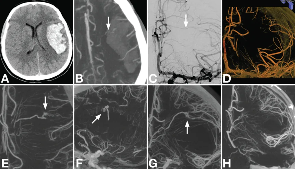
This report describes 3 patients with brain bleeding who were studied with cone-beam CT angiography, a high-resolution 3D scan done in the angiography room, along with standard imaging. In all 3, it showed the detailed structure of an AVM where other tests had shown nothing or only a hint of a draining vein. In 2 patients the AVM was then fully removed during surgery, and in the third it allowed precise targeting with Gamma Knife radiosurgery.
The authors recommend considering this scan when a hidden AVM is suspected.
A plain-language summary for patients and families. It is not medical advice, and individual results vary.
Abstract
Object
Ruptured arteriovenous malformations (AVMs) are a frequent cause of intracerebral hemorrhage (ICH). In some cases, compression from the associated hematoma in the acute setting can partially or completely occlude an AVM, making it invisible on conventional angiography techniques. The authors report on the successful use of cone-beam CT angiography (CBCT-A) to precisely identify the underlying angioarchitecture of ruptured AVMs that are not visible on conventional angiography.
Methods
Three patients presented with ICH for which they underwent examination with CBCT-A in addition to digital subtraction angiography and other imaging modalities, including MR angiography and CT angiography. All patients underwent surgical evacuation due to mass effect from the hematoma. Clinical history, imaging studies, and surgical records were reviewed. Hematoma volumes were calculated.
Results
In all 3 cases, CBCT-A demonstrated detailed anatomy of an AVM where no lesion or just a suggestion of a draining vein had been seen with other imaging modalities. Magnetic resonance imaging demonstrated enhancement in 1 patient; CT angiography demonstrated a draining vein in 1 patient; 2D digital subtraction angiography and 3D rotational angiography demonstrated a suggestion of a draining vein in 2 cases and no finding in the third. In the 2 patients in whom CBCT-A was performed prior to surgery, the demonstrated AVM was successfully resected without evidence of a residual lesion. In the third patient, CBCT-A allowed precise targeting of the AVM nidus using Gamma Knife radiosurgery.
Conclusions
Cone-beam CT angiography should be considered in the evaluation and subsequent treatment of ICH due to ruptured AVMs. In cases in which the associated hematoma compresses the AVM nidus, CBCT-A can have higher sensitivity and anatomical accuracy than traditional angiographic modalities, including digital subtraction angiography.
Abstract as indexed in PubMed (PMID 23683076). Copyright the publisher.
Cite this paper
Rahal JP, Malek AM. Benefit of cone-beam computed tomography angiography in acute management of angiographically undetectable ruptured arteriovenous malformations. J Neurosurg 119(4):1015-20. 2013. doi:10.3171/2013.4.JNS1390 PMID: 23683076.