2008 · Devices & clinical
Stent-mediated wedging of a calcific embolus to recanalize an occluded middle cerebral artery: technical case report
Schirmer CM, Thaler DE, Malek AM.
Neurosurgery 63(1):E180-1; discussion E181, 2008.
PMID 18728560 · DOI 10.1227/01.NEU.0000335088.85470.FC
In plain language
Sometimes a piece of calcium, rather than an ordinary blood clot, breaks loose and blocks a brain artery. These calcific emboli do not respond to clot-dissolving drugs and tend to break apart, making them hard to treat.
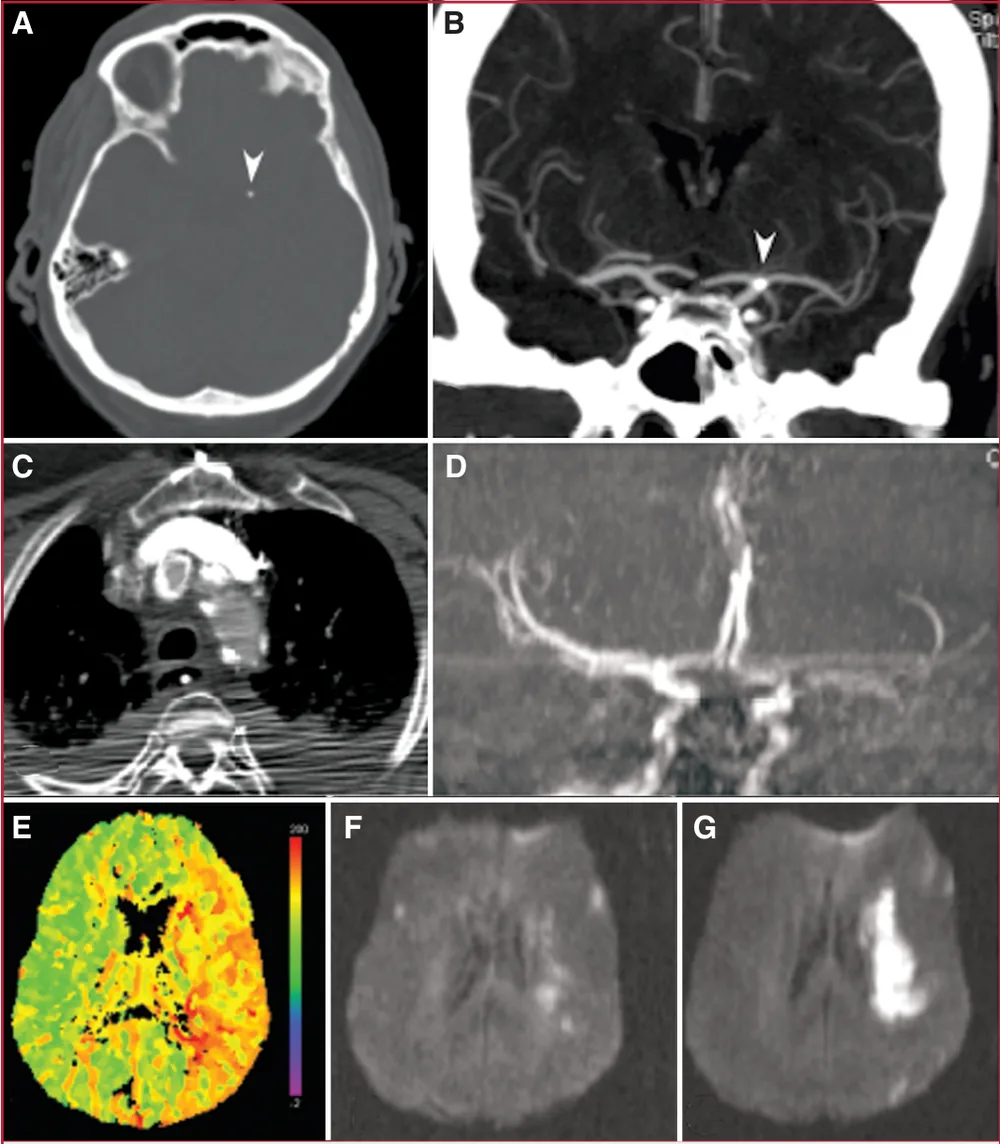
This report describes a 75-year-old woman who developed trouble speaking and weakness on one side after a heart catheterization, caused by a calcium fragment nearly blocking her left middle cerebral artery. She worsened despite 6 days of medical treatment. Attempts to pull out the fragment with standard retrieval devices failed and caused it to block the artery completely. The team then used a balloon-mounted stent to press the fragment against the artery wall, which reopened the artery and restored blood flow.
A plain-language summary for patients and families. It is not medical advice, and individual results vary.
Abstract
Objective
Calcific cerebral emboli are rare yet pose a difficult therapeutic challenge because of their lack of response to thrombolytic therapy and their propensity for fragmentation.
Clinical presentation
A 75-year-old woman developed aphasia and hemiparesis after cardiac catheterization from a near-occlusive calcific embolus to the left middle cerebral artery origin. The calcific embolus was shown to be hemodynamically limiting by computed tomographic perfusion and diffusion-weighted magnetic resonance imaging.
Intervention
The patient was treated medically for 6 days with anticoagulation, antiplatelet, and hypertensive therapies but continued to deteriorate. A decision was made to attempt retrieval of the calcific embolus. Conventional intracranial mechanical clot and coil retrieval systems were used initially but were unsuccessful and caused migration of the calcific embolus, leading to complete occlusion of the left middle cerebral artery origin. At this point, the decision was made to wedge the calcific embolus against the vessel wall. A balloon-mounted coronary stent was used to achieve this successfully, resulting in recanalization of the middle cerebral artery trunk and flow restoration.
Conclusion
Symptomatic calcific emboli, which are poorly responsive to pharmacological therapy and difficult to recover using conventional retrieval systems, may be treated by immobilization against the vessel wall using a balloon-mounted stent.
Abstract as indexed in PubMed (PMID 18728560). Copyright the publisher.
Cite this paper
Schirmer CM, Thaler DE, Malek AM. Stent-mediated wedging of a calcific embolus to recanalize an occluded middle cerebral artery: technical case report. Neurosurgery 63(1):E180-1; discussion E181. 2008. doi:10.1227/01.NEU.0000335088.85470.FC PMID: 18728560.