2025 · Hemodynamics & imaging
A Novel Application of Radiomics Analysis for the Evaluation of Centroid Radii Model Texture on the Surface of Cerebral Aneurysms
Lauric A, Malek AM.
Stroke Vasc Interv Neurol 5(6):e001909, 2025.
PMID 41608704 · DOI 10.1161/SVIN.125.001909 · PMCID PMC12697587
Narrated summary
Figures from the open access article (CC BY-NC 4.0).
Read the transcript
Here's a short summary of our recent study from our laboratory at Tufts Medical Center, led by Dr. Alexandra Lauric. I'm Adel Malek. Brain aneurysms are common, and most never rupture. Deciding which ones to treat is one of the hardest calls in our field. Size helps, but many small aneurysms rupture, and many large ones never do. So we look for other clues in the aneurysm's shape. One clue is irregularity. The centroid radii model measures the distance from every point on the aneurysm's surface to its center.
A smooth, round aneurysm has nearly equal distances everywhere. A lobulated one, with blebs, has peaks and valleys. We projected those distances onto a sphere, unrolled the sphere into a flat image, and analyzed that image with radiomics: software that measures texture and pattern, much as it's used to study tumors on scans. That gave us 93 features for each aneurysm. We studied 187 aneurysms imaged with three-dimensional angiography.
49 of them had ruptured. Ruptured aneurysms looked different. Their maps were more complex and uneven, with small, localized peaks, like blebs, scattered over wide, low areas. Unruptured aneurysms looked smoother and more uniform. A model built from five texture features separated ruptured from unruptured aneurysms with an area under the curve of 0.86, compared with 0.82 for the standard size and shape measures. Texture analysis of the aneurysm surface may add to how we assess rupture risk.
Because this study looked back at aneurysms that had already ruptured, the next step is testing whether it predicts future rupture.
In plain language
Shape can help reveal which brain aneurysms have ruptured. One measure, the centroid radii model, records the distance from each point on the aneurysm surface to its center. This study applied radiomics, a method that analyzes texture and pattern in images, to maps of these distances.
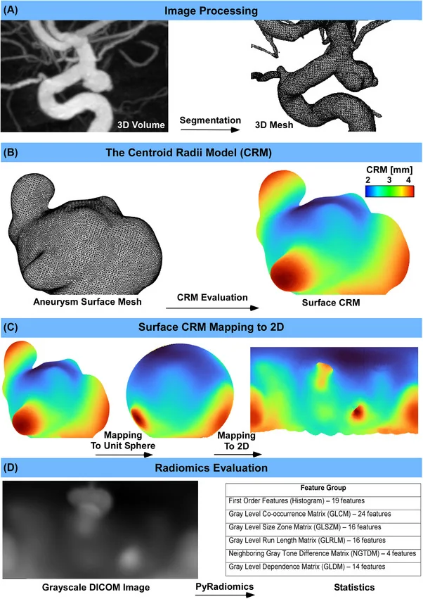
In 187 aneurysms, 49 of which had ruptured, the distance maps were projected onto a sphere and turned into grayscale images. Ruptured aneurysms showed more complex, uneven patterns. A model built from these features separated ruptured from unruptured aneurysms with an area under the curve of 0.86 (1.0 would be perfect), compared with 0.82 for standard size and shape measures.
This new use of radiomics may eventually help with assessing aneurysm risk.
A plain-language summary for patients and families. It is not medical advice, and individual results vary.
Abstract
Background
The centroid radii model (CRM)-defined as the distance from aneurysm surface points to the centroid-has shown promise in distinguishing ruptured from unruptured intracranial aneurysms by capturing morphological characteristics. We propose a novel application of radiomics for analyzing CRM texture and patterns, extending radiomics beyond medical imaging-based analysis.
Methods
Three-dimensional rotational angiographic volumes from 187 aneurysms (49 ruptured) were analyzed. Aneurysm surfaces were segmented and converted to uniform triangular meshes. Established size and shape metrics (size, height, aspect ratio, height/width, size ratio, and nonsphericity) and CRM values were computed. CRM data were projected onto a unit sphere and mapped to grayscale images for radiomics analysis. Univariate and multivariate analyses were used to evaluate the established features and 93 radiomics features for the accuracy of rupture status discrimination.
Results
Ruptured aneurysms exhibited greater CRM texture complexity and heterogeneity, with higher entropy, variance, contrast, and gray-level nonuniformity. These aneurysms had localized high CRM intensities and widespread low CRM regions, as well as reduced pattern uniformity. All established morphological features were significantly elevated in ruptured aneurysms. Multivariate regression using radiomics features resulted in an area under the curve of 0.86 (specificity 0.81, sensitivity 0.78), compared with conventional features (area under the curve 0.82, specificity 0.75, sensitivity 0.81).
Conclusion
Radiomics-based histogram and texture analysis of surface CRM offers strong rupture status discrimination power, which compares favorably with established size and shape features. This novel use of radiomics on surface-based features provides additional insight into the characteristics of ruptured aneurysms and may have potential utility in risk stratification.
Abstract as indexed in PubMed (PMID 41608704). Copyright the publisher.
Cite this paper
Lauric A, Malek AM. A Novel Application of Radiomics Analysis for the Evaluation of Centroid Radii Model Texture on the Surface of Cerebral Aneurysms. Stroke Vasc Interv Neurol 5(6):e001909. 2025. doi:10.1161/SVIN.125.001909 PMID: 41608704.