1993 · Mechanobiology
Fluid shear stress differentially modulates expression of genes encoding basic fibroblast growth factor and platelet-derived growth factor B chain in vascular endothelium
Malek AM, Gibbons GH, Dzau VJ, Izumo S.
J Clin Invest 92(4):2013-21, 1993.
PMID 8408655 · DOI 10.1172/JCI116796 · PMCID PMC288369
In plain language
Shear stress, the rubbing force of blood flow on the vessel lining, helps shape vessel structure through its effects on endothelial cells, the cells that line blood vessels. This laboratory study examined two growth factors these cells make, PDGF-B and bFGF, signals that can promote cell growth.
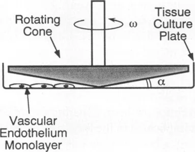
Cow aortic endothelial cells were exposed to flow in a purpose-built cone-plate device. Higher force (36 dyn/cm2) aligned the cells with the flow more strongly than moderate force (15 dyn/cm2). PDGF-B mRNA fell 3.9-fold and 4.2-fold at these two levels by 9 hours. In contrast, bFGF rose only mildly and briefly at 15 dyn/cm2 but increased 4.8-fold at 36 dyn/cm2 after 6 hours, remaining 2.9-fold higher at 9 hours. Pulsing and turbulent flow acted like steady flow, and stretching had no significant effect.
Flow therefore regulates these two growth factor genes differently.
A plain-language summary for patients and families. It is not medical advice, and individual results vary.
Abstract
Fluid shear stress has been shown to be an important regulator of vascular structure and function through its effect on the endothelial cell. We have explored the effect of shear stress on the expression of the heparin-binding growth factors platelet-derived growth factor B chain (PDGF-B) and basic fibroblast growth factor (bFGF) in bovine aortic endothelial cells using a purpose-built cone-plate viscometer. Using morphometric analysis, we have mimicked the endothelial cell shape changes encountered in vivo in response to shear stress and correlated these with changes in gene expression. Steady laminar shear stress of 15 and 36 dyn/cm2 both resulted in endothelial cell shape change, but the higher shear stress induced greater and more uniform alignment in the direction of flow and nuclear protrusion after 24 h. Steady laminar shear stress of both 15 and 36 dyn/cm2 induced a significant 3.9- and 4.2-fold decrease, respectively, in PDGF-B mRNA at 9 h. In contrast, steady laminar shear of 15 dyn/cm2 induced a mild and transient 1.5-fold increase in bFGF mRNA while shear of 36 dyn/cm2 induced a significant 4.8-fold increase at 6 h of shear which remained at 2.9-fold at 9 h. Pulsatile and turbulent shear stress showed the same effect as steady laminar shear stress (all at 15 dyn/cm2 time-average magnitude) on PDGF-B and bFGF mRNA content. Cyclic stretch (20% strain, 20/min) of cells grown on silicone substrate did not significantly affect either PDGF-B or bFGF mRNA levels. These results suggest that expression of each peptide growth factor gene is differentially regulated by fluid shear stress in the vascular endothelial cell. These results may have implications on vascular structure and function in response to hemodynamic forces and present a model for the study of transduction of mechanical stimuli into altered gene expression.
Abstract as indexed in PubMed (PMID 8408655). Copyright the publisher.
Cite this paper
Malek AM, Gibbons GH, Dzau VJ, Izumo S. Fluid shear stress differentially modulates expression of genes encoding basic fibroblast growth factor and platelet-derived growth factor B chain in vascular endothelium. J Clin Invest 92(4):2013-21. 1993. doi:10.1172/JCI116796 PMID: 8408655.