1997 · Mechanobiology
Regulation of endothelin-1 gene expression by cell shape and the microfilament network in vascular endothelium
Malek AM, Lee IW, Alper SL, Izumo S.
Am J Physiol 273(5):C1764-74, 1997.
PMID 9374665 · DOI 10.1152/ajpcell.1997.273.5.C1764
In plain language
Endothelin-1 is a vessel-tightening substance made by endothelial cells, the cells that line blood vessels, and its production responds to flow and stretch. This study tested whether simply changing the shape of these cells could control the endothelin-1 gene.
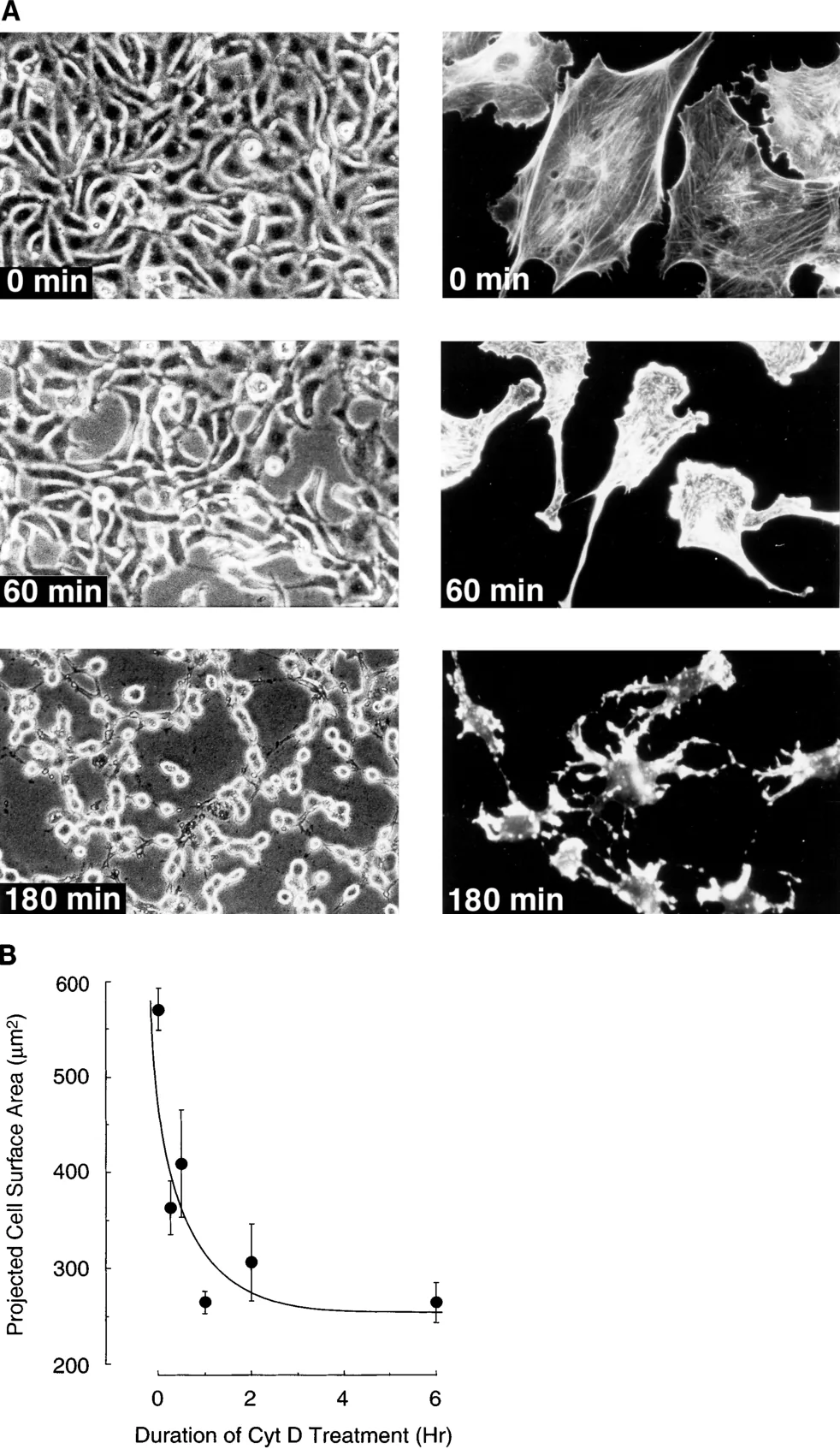
In cow aortic endothelial cells, a drug that breaks down actin fibers (part of the cell's internal scaffolding) made the cells round up and lowered endothelin-1 mRNA. Several other drugs that changed cell shape had the same effect. Cells grown on surfaces that let them spread less built fewer actin fibers, secreted 60% less endothelin-1 and had 94% less endothelin-1 mRNA. This was not explained by reduced contact between cells.
Cell shape and the actin network appear to be strong controllers of this gene, acting after the gene is copied into mRNA.
A plain-language summary for patients and families. It is not medical advice, and individual results vary.
Abstract
Endothelial synthesis and release of endothelin-1 (ET-1) are exquisitely regulated by external shear and strain. We tested the hypothesis that manipulation of endothelial cell shape can regulate ET-1 gene expression. Treatment of bovine aortic endothelial cell (BAEC) monolayers with cytochalasin D disrupted F-actin and induced cell retraction and rounding, in parallel with time- and dose-dependent specific decreases in ET-1 mRNA levels. Treatments with forskolin, phorbol 12-myristate 13-acetate, staurosporine, and genistein also induced cell shape change and decreased F-actin staining and ET-1 mRNA levels. BAEC plated onto nonadhesive petri dishes coated with decreasing concentrations of synthetic RGD polymer showed RGD dose-dependent decreases in cell spreading and in F-actin microfilament elaboration. These changes were specifically accompanied by decreases in ET-1 peptide secretion (60%) and, via posttranscriptional mechanisms, ET-1 mRNA (94%) and were not due to decreased cell-cell contact. We conclude that the shape and microfilament network of endothelial cells are potent posttranscriptional regulators of ET-1 gene expression.
Abstract as indexed in PubMed (PMID 9374665). Copyright the publisher.
Cite this paper
Malek AM, Lee IW, Alper SL, Izumo S. Regulation of endothelin-1 gene expression by cell shape and the microfilament network in vascular endothelium. Am J Physiol 273(5):C1764-74. 1997. doi:10.1152/ajpcell.1997.273.5.C1764 PMID: 9374665.