2026 · Devices & clinical
Development, image interpretation, clinical experience, and applications of optical coherence tomography in neurointerventional surgery
Lopes D, Hanel RA, Anagnostakou V, Cancelliere NM, Fiehler J, Lylyk I, Lylyk PN, Rouchaud A, Forestier G, Malek AM, Vakharia K, Mokin M, Ogilvy CS, Osborn E, Bezerra H, Uemura S, Siddiqui A, Andersson T, Gounis MJ, Lylyk P, Pereira VM, Ughi GJ.
J Neurointerv Surg 18(8):1796-1811, 2026.
PMID 41720623 · DOI 10.1136/jnis-2025-024359
In plain language
Angiography, the standard X-ray movie of blood vessels, shows the channel that blood flows through but not the vessel wall itself, or exactly how a device sits against it. Optical coherence tomography (OCT) uses light from a tiny probe inside the artery to create very detailed 3D pictures, and heart specialists already rely on it during complex stent procedures.
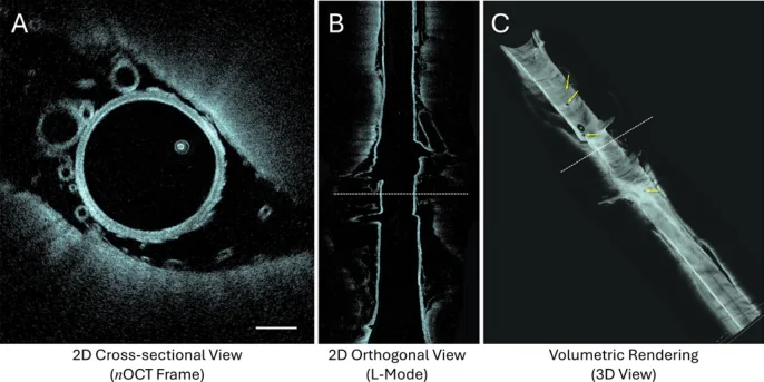
In this review, an international group of experts explains how a new OCT system designed for the narrow, winding arteries of the brain works, how to read its images, and how it may help doctors understand vessel disease and check stents and other devices during treatment.
A plain-language summary for patients and families. It is not medical advice, and individual results vary.
Abstract
Angiography remains the standard imaging modality for evaluating vascular disease and guiding endovascular interventions; however, its limited spatial resolution restricts visualization of vessel wall pathology and therapeutic devices. Over the past decade, intravascular optical coherence tomography (OCT) has emerged as a transformative tool in coronary artery disease. OCT provides three-dimensional, high-resolution imaging of coronary arteries, enabling assessment of vessel wall microstructure, plaque composition, lumen geometry, and stent deployment. This level of detail has advanced the understanding of coronary pathology and contributed to improved outcomes in percutaneous coronary interventions. Major cardiology societies now recommend the use of intravascular imaging, including OCT, for complex coronary interventions with level 1A evidence.OCT is now expanding beyond the coronary vasculature, with growing interest in its potential role in neurointerventional surgery. While limited use of OCT has been reported in the neurovasculature, the characteristics of a coronary OCT device prevent routine use in intracranial arteries. A new generation of intravascular OCT technology has been developed for the tortuous and complex anatomy of cerebral vessels. Early clinical experience has investigated its safety, feasibility, and utility for intracranial vascular imaging. Neuro-dedicated OCT technology could represent a significant advancement in cerebrovascular imaging, offering insights beyond the capabilities of current modalities, with the potential to improve current understanding of neurovascular pathology and refine diagnostic and therapeutic strategies. Successful translation of OCT into neurointervention, however, also requires establishing principles for image interpretation to assess neurovascular disease and therapeutic devices. This article aims to provide guidance for interpreting OCT imaging of intracranial arteries. In addition, it reviews applications of OCT in neurointervention and describes the development of neuro-specific OCT technology.
Abstract as indexed in PubMed (PMID 41720623). Copyright the publisher.
Cite this paper
Lopes D, Hanel RA, Anagnostakou V, Cancelliere NM, Fiehler J, Lylyk I, Lylyk PN, Rouchaud A, Forestier G, Malek AM, Vakharia K, Mokin M, Ogilvy CS, Osborn E, Bezerra H, Uemura S, Siddiqui A, Andersson T, Gounis MJ, Lylyk P, Pereira VM, Ughi GJ. Development, image interpretation, clinical experience, and applications of optical coherence tomography in neurointerventional surgery. J Neurointerv Surg 18(8):1796-1811. 2026. doi:10.1136/jnis-2025-024359 PMID: 41720623.