2019 · Devices & clinical
Anatomical characterization of the inferior petrosal sinus and adjacent cerebellopontine angle cistern for development of an endovascular transdural cerebrospinal fluid shunt
Heilman CB, Basil GW, Beneduce BM, Malek AM.
J Neurointerv Surg 11(6):598-602, 2019.
PMID 30626626 · DOI 10.1136/neurintsurg-2018-014445
In plain language
In communicating hydrocephalus, cerebrospinal fluid, the clear fluid that cushions the brain, builds up because it is not absorbed properly into the bloodstream. The authors are developing a shunt that could be delivered through a vein by catheter to create a new drainage path, and such a device needs a safe landing site.
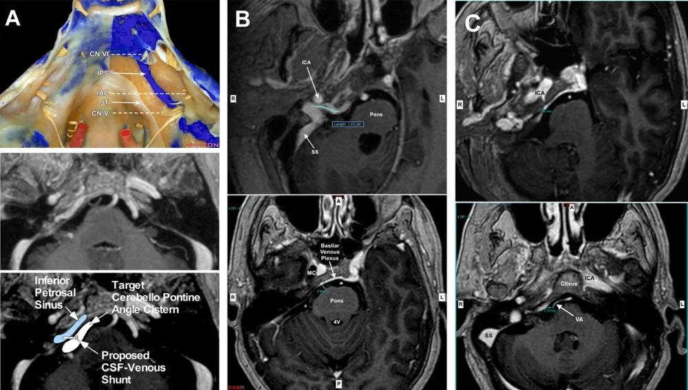
Brain MRI scans from 36 patients aged 20 to 80 years were measured in three dimensions to study the inferior petrosal sinus, a vein at the skull base, and the fluid filled space beside it. Average vein diameter ranged from 2.27 millimeters to 3.31 millimeters, with more than 90 percent of measurements larger than 1.46 millimeters, and the neighboring fluid space measured 3.86 millimeters to 7.39 millimeters deep.
The measurements suggest there is enough room for a catheter delivered shunt at this location.
A plain-language summary for patients and families. It is not medical advice, and individual results vary.
Abstract
Background and purpose
We evaluated the inferior petrosal sinus (IPS) and adjacent cerebellopontine angle (CPA) cistern as a potential implantation site for a novel venous endovascular transdural CSF shunt concept to treat communicating hydrocephalus. We analyzed the dimensions of the IPS, CPA cistern, and distances to adjacent neurovascular structures.
Materials and methods
Gadolinium enhanced T1 weighted brain MRI datasets of 36 randomly selected patients, aged 20-80 years, were analyzed with three-dimensional multiplanar reconstruction to measure IPS diameter and length, CPA cistern depth, and IPS proximity to the vertebrobasilar arteries and brainstem. Statistical analysis was used to assess gender, sidedness, and age dependence.
Results
Mean IPS diameter ranged from 2.27 mm to 3.31 mm at three axial levels, with >90% larger than 1.46 mm. CPA cistern adjacent to the IPS exhibited a mean depth of 3.86 mm to 7.39 mm between the dura and brainstem at corresponding axial levels. There was no side dependence except for a longer distance from the IPS to the basilar artery on the left compared with the right (9.72 vs 7.28, P<0.019). Linear regression analysis showed that the distance from the IPS to the brainstem was statistically significantly increased with age (P<0.0002) and was greater in men, with little side variation (P=0.524).
Conclusion
Our results demonstrate sufficient CSF CPA cisternal space adjacent to the IPS and support the feasibility of an endovascular catheter delivered transdural implantable shunt. Such a device could serve to mimic the function of the arachnoid granulation by establishing a regulated path for CSF flow from the intracranial subarachnoid space to the venous system and provide a treatment for communicating hydrocephalus.
Abstract as indexed in PubMed (PMID 30626626). Copyright the publisher.
Cite this paper
Heilman CB, Basil GW, Beneduce BM, Malek AM. Anatomical characterization of the inferior petrosal sinus and adjacent cerebellopontine angle cistern for development of an endovascular transdural cerebrospinal fluid shunt. J Neurointerv Surg 11(6):598-602. 2019. doi:10.1136/neurintsurg-2018-014445 PMID: 30626626.